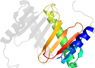
Lineage for d1y4oa1 (1y4o A:10-104)
- Root: SCOPe 2.07

Class d: Alpha and beta proteins (a+b) [53931] (388 folds) 
Fold d.110: Profilin-like [55769] (10 superfamilies)
core: 2 alpha-helices and 5-stranded antiparallel sheet: order 21543; 3 layers: alpha/beta/alpha
Superfamily d.110.7: Roadblock/LC7 domain [103196] (2 families) 
alpha-beta(2)-alpha-beta(3)-alpha; structurally most similar to the SNARE-like superfamily with a circular permutation of the terminal helices
Family d.110.7.1: Roadblock/LC7 domain [103197] (5 proteins)
Pfam PF03259
Protein Dynein light chain 2A, cytoplasmic [118074] (2 species) 
Species Mouse (Mus musculus) [TaxId:10090] [160682] (1 PDB entry)
Uniprot P62627 1-95
Domain d1y4oa1: 1y4o A:10-104 [145901]
Other proteins in same PDB: d1y4oa2, d1y4ob3
Details for d1y4oa1
PDB Entry: 1y4o (more details)
PDB Description: solution structure of a mouse cytoplasmic roadblock/lc7 dynein light chain
PDB Compounds: (A:) Dynein light chain 2A, cytoplasmicSCOPe Domain Sequences for d1y4oa1:
Sequence; same for both SEQRES and ATOM records: (download)
>d1y4oa1 d.110.7.1 (A:10-104) Dynein light chain 2A, cytoplasmic {Mouse (Mus musculus) [TaxId: 10090]}
aeveetlkrlqsqkgvqgiivvntegipikstmdnptttqyanlmhnfilkarstvreid
pqndltflrirskkneimvapdkdyfliviqnpte
SCOPe Domain Coordinates for d1y4oa1:
Click to download the PDB-style file with coordinates for d1y4oa1.
(The format of our PDB-style files is described here.)
(The format of our PDB-style files is described here.)
Timeline for d1y4oa1:
- d1y4oa1 first appeared in SCOP 1.75
- d1y4oa1 appears in SCOPe 2.06
- d1y4oa1 appears in the current release, SCOPe 2.08

 View in 3D


