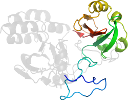
Lineage for d3s7oa2 (3s7o A:135-323)
- Root: SCOPe 2.04

Class d: Alpha and beta proteins (a+b) [53931] (380 folds) 
Fold d.110: Profilin-like [55769] (10 superfamilies)
core: 2 alpha-helices and 5-stranded antiparallel sheet: order 21543; 3 layers: alpha/beta/alpha
Superfamily d.110.2: GAF domain-like [55781] (5 families) 
alpha(2)-beta(3)-alpha-beta(3)-alpha; antiparallel beta-sheet: order 321654
Family d.110.2.1: GAF domain [55782] (8 proteins) Protein automated matches [227076] (2 species)
not a true proteinSpecies Deinococcus radiodurans [TaxId:1299] [226274] (5 PDB entries) 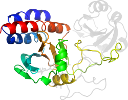
Domain d3s7oa2: 3s7o A:135-323 [216231]
Other proteins in same PDB: d3s7oa1
automated match to d1ztua1
complexed with gol, lbv
Details for d3s7oa2
PDB Entry: 3s7o (more details), 1.24 Å
PDB Description: Crystal Structure of the Infrared Fluorescent D207H variant of Deinococcus Bacteriophytochrome chromophore binding domain at 1.24 angstrom resolution
PDB Compounds: (A:) BacteriophytochromeSCOPe Domain Sequences for d3s7oa2:
Sequence; same for both SEQRES and ATOM records: (download)
>d3s7oa2 d.110.2.1 (A:135-323) automated matches {Deinococcus radiodurans [TaxId: 1299]}
tgphalrnamfalesapnlralaevatqtvreltgfdrvmlykfapdatgeviaearreg
lhaflghrfpashipaqaralytrhllrltadtraaavpldpvlnpqtnaptplggavlr
atspmhmqylrnmgvgsslsvsvvvggqlwgliachhqtpyvlppdlrttleslgrllsl
qvqvkeale
SCOPe Domain Coordinates for d3s7oa2:
Click to download the PDB-style file with coordinates for d3s7oa2.
(The format of our PDB-style files is described here.)
(The format of our PDB-style files is described here.)
Timeline for d3s7oa2:
- d3s7oa2 first appeared in SCOPe 2.03
- d3s7oa2 appears in SCOPe 2.05
- d3s7oa2 appears in the current release, SCOPe 2.08

 View in 3D