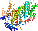
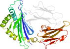
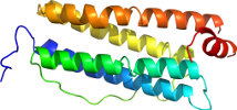
Lineage for d2vhnw1 (2vhn W:1-84)
- Root: SCOPe 2.08

Class b: All beta proteins [48724] (180 folds) 
Fold b.84: Barrel-sandwich hybrid [51229] (5 superfamilies)
sandwich of half-barrel shaped beta-sheets
Superfamily b.84.4: Ribosomal L27 protein-like [110324] (3 families) 
rudiment single hybrid fold with a permuted topology
Family b.84.4.1: Ribosomal L27 protein [110325] (1 protein)
Pfam PF01016Protein Ribosomal protein L27 [110326] (3 species) 
Species Escherichia coli [TaxId:562] [159322] (29 PDB entries)
Uniprot P0A7L8 1-84
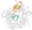
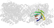
Domain d2vhnw1: 2vhn W:1-84 [153118]
Other proteins in same PDB: d2vhn01, d2vhn11, d2vhn31, d2vhn41, d2vhnc1, d2vhnc2, d2vhnd1, d2vhne1, d2vhnf1, d2vhng1, d2vhng2, d2vhnh1, d2vhnh2, d2vhni1, d2vhni2, d2vhnj1, d2vhnk1, d2vhnl1, d2vhnm1, d2vhnn1, d2vhno1, d2vhnp1, d2vhnq1, d2vhnr1, d2vhns1, d2vhnt1, d2vhnu1, d2vhnv1, d2vhnx1, d2vhny1, d2vhnz1
protein/RNA complex; complexed with mg
protein/RNA complex; complexed with mg
Details for d2vhnw1
PDB Entry: 2vhn (more details), 3.74 Å
PDB Description: Structure of PDF binding helix in complex with the ribosome. (Structure 2 of 4)
PDB Compounds: (W:) 50S ribosomal protein L27SCOPe Domain Sequences for d2vhnw1:
Sequence; same for both SEQRES and ATOM records: (download)
>d2vhnw1 b.84.4.1 (W:1-84) Ribosomal protein L27 {Escherichia coli [TaxId: 562]}
ahkkaggstrngrdseakrlgvkrfggesvlagsiivrqrgtkfhaganvgcgrdhtlfa
kadgkvkfevkgpknrkfisieae
SCOPe Domain Coordinates for d2vhnw1:
Click to download the PDB-style file with coordinates for d2vhnw1.
(The format of our PDB-style files is described here.)
(The format of our PDB-style files is described here.)
Timeline for d2vhnw1:
- d2vhnw1 first appeared in SCOP 1.75
- d2vhnw1 appears in SCOPe 2.07
 View in 3D View in 3DDomains from other chains: (mouse over for more information) d2vhn01, d2vhn11, d2vhn31, d2vhn41, d2vhnc1, d2vhnc2, d2vhnd1, d2vhne1, d2vhnf1, d2vhng1, d2vhng2, d2vhnh1, d2vhnh2, d2vhni1, d2vhni2, d2vhnj1, d2vhnk1, d2vhnl1, d2vhnm1, d2vhnn1, d2vhno1, d2vhnp1, d2vhnq1, d2vhnr1, d2vhns1, d2vhnt1, d2vhnu1, d2vhnv1, d2vhnx1, d2vhny1, d2vhnz1 |