Lineage for d1vs6o1 (1vs6 O:1-117)
- Root: SCOPe 2.08

Class c: Alpha and beta proteins (a/b) [51349] (148 folds) 
Fold c.55: Ribonuclease H-like motif [53066] (7 superfamilies)
3 layers: a/b/a; mixed beta-sheet of 5 strands, order 32145; strand 2 is antiparallel to the rest
Superfamily c.55.4: Translational machinery components [53137] (3 families) 
Family c.55.4.1: Ribosomal protein L18 and S11 [53138] (2 proteins) Protein Ribosomal protein L18 (L18p) [53139] (5 species) 
Species Escherichia coli [TaxId:562] [159642] (29 PDB entries)
Uniprot P0C018 1-117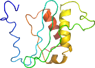
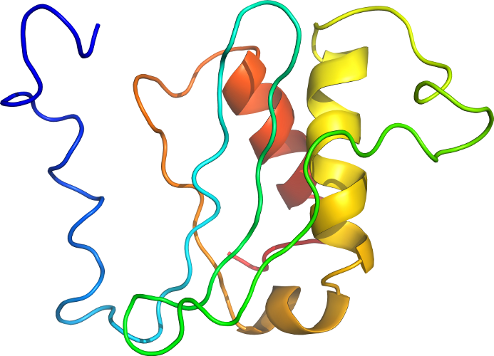
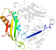
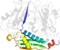
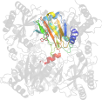
Domain d1vs6o1: 1vs6 O:1-117 [144470]
Other proteins in same PDB: d1vs601, d1vs611, d1vs621, d1vs631, d1vs641, d1vs6c1, d1vs6c2, d1vs6d1, d1vs6e1, d1vs6f1, d1vs6g1, d1vs6g2, d1vs6h1, d1vs6h2, d1vs6i1, d1vs6i2, d1vs6j1, d1vs6k1, d1vs6l1, d1vs6m1, d1vs6n1, d1vs6p1, d1vs6q1, d1vs6r1, d1vs6s1, d1vs6t1, d1vs6u1, d1vs6v1, d1vs6w1, d1vs6x1, d1vs6y1, d1vs6z1
complexed with mg
complexed with mg
has additional insertions and/or extensions that are not grouped together
Details for d1vs6o1
PDB Entry: 1vs6 (more details), 3.46 Å
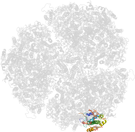
PDB Description: Crystal structure of the bacterial ribosome from escherichia coli in complex with the antibiotic kasugamyin at 3.5A resolution. this file contains the 50s subunit of one 70s ribosome. the entire crystal structure contains two 70s ribosomes and is described in remark 400.
PDB Compounds: (O:) 50S ribosomal protein L18SCOPe Domain Sequences for d1vs6o1:
Sequence; same for both SEQRES and ATOM records: (download)
>d1vs6o1 c.55.4.1 (O:1-117) Ribosomal protein L18 (L18p) {Escherichia coli [TaxId: 562]}
mdkksarirratrarrklqelgatrlvvhrtprhiyaqviapngsevlvaastvekaiae
qlkytgnkdaaaavgkavaeralekgikdvsfdrsgfqyhgrvqaladaareaglqf
SCOPe Domain Coordinates for d1vs6o1:
Click to download the PDB-style file with coordinates for d1vs6o1.
(The format of our PDB-style files is described here.)
(The format of our PDB-style files is described here.)
Timeline for d1vs6o1:
- d1vs6o1 first appeared in SCOP 1.75
- d1vs6o1 appears in SCOPe 2.07
 View in 3D View in 3DDomains from other chains: (mouse over for more information) d1vs601, d1vs611, d1vs621, d1vs631, d1vs641, d1vs6c1, d1vs6c2, d1vs6d1, d1vs6e1, d1vs6f1, d1vs6g1, d1vs6g2, d1vs6h1, d1vs6h2, d1vs6i1, d1vs6i2, d1vs6j1, d1vs6k1, d1vs6l1, d1vs6m1, d1vs6n1, d1vs6p1, d1vs6q1, d1vs6r1, d1vs6s1, d1vs6t1, d1vs6u1, d1vs6v1, d1vs6w1, d1vs6x1, d1vs6y1, d1vs6z1 |