Lineage for d3ci3a1 (3ci3 A:2-188)
- Root: SCOPe 2.06

Class a: All alpha proteins [46456] (289 folds) 
Fold a.25: Ferritin-like [47239] (6 superfamilies)
core: 4 helices; bundle, closed, left-handed twist; 1 crossover connection
Superfamily a.25.2: Cobalamin adenosyltransferase-like [89028] (3 families) 
crossover loop goes across a different side of the 4-helical bundle; no internal metal-binding siteFamily a.25.2.0: automated matches [191442] (1 protein)
not a true familyProtein automated matches [190652] (6 species)
not a true proteinSpecies Lactobacillus reuteri [TaxId:1598] [188444] (9 PDB entries) 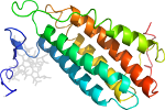
Domain d3ci3a1: 3ci3 A:2-188 [173246]
Other proteins in same PDB: d3ci3a2
automated match to d1rtyb_
complexed with 3po, 5ad, b12, mg, na
Details for d3ci3a1
PDB Entry: 3ci3 (more details), 1.11 Å
PDB Description: Structure of the PduO-type ATP:co(I)rrinoid adenosyltransferase from Lactobacillus reuteri complexed with partial adenosylcobalamin and PPPi
PDB Compounds: (A:) Cobalamin adenosyltransferase PduO-like proteinSCOPe Domain Sequences for d3ci3a1:
Sequence; same for both SEQRES and ATOM records: (download)
>d3ci3a1 a.25.2.0 (A:2-188) automated matches {Lactobacillus reuteri [TaxId: 1598]}
kiytkngdkgqtriigkqilykndprvaaygevdelnswvgytkslinshtqvlsnelee
iqqllfdcghdlatpadderhsfkfkqeqptvwleekidnytqvvpavkkfilpggtqla
salhvartitrraerqivqlmreeqinqdvlifinrlsdyffaaaryanyleqqpdmlyr
nskdvfr
SCOPe Domain Coordinates for d3ci3a1:
Click to download the PDB-style file with coordinates for d3ci3a1.
(The format of our PDB-style files is described here.)
(The format of our PDB-style files is described here.)
Timeline for d3ci3a1:
- d3ci3a1 first appeared in SCOPe 2.01, called d3ci3a_
- d3ci3a1 was called d3ci3a_ in SCOPe 2.05
- d3ci3a1 appears in SCOPe 2.07
- d3ci3a1 appears in the current release, SCOPe 2.08

 View in 3D
