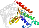
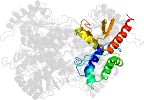
Lineage for d3pqfd1 (3pqf D:3-147)
- Root: SCOPe 2.06

Class c: Alpha and beta proteins (a/b) [51349] (148 folds) 
Fold c.2: NAD(P)-binding Rossmann-fold domains [51734] (1 superfamily)
core: 3 layers, a/b/a; parallel beta-sheet of 6 strands, order 321456
The nucleotide-binding modes of this and the next two folds/superfamilies are similarSuperfamily c.2.1: NAD(P)-binding Rossmann-fold domains [51735] (13 families) 

Family c.2.1.0: automated matches [191313] (1 protein)
not a true familyProtein automated matches [190069] (239 species)
not a true protein
Species Bacillus subtilis [TaxId:1423] [196388] (11 PDB entries) 
Domain d3pqfd1: 3pqf D:3-147 [214931]
Other proteins in same PDB: d3pqfa2, d3pqfb2, d3pqfc2, d3pqfd2
automated match to d1llca1
complexed with nad; mutant
Details for d3pqfd1
PDB Entry: 3pqf (more details), 2.49 Å
PDB Description: Crystal structure of L-lactate dehydrogenase from Bacillus subtilis mutation H171C complexed with NAD+
PDB Compounds: (D:) l-lactate dehydrogenaseSCOPe Domain Sequences for d3pqfd1:
Sequence; same for both SEQRES and ATOM records: (download)
>d3pqfd1 c.2.1.0 (D:3-147) automated matches {Bacillus subtilis [TaxId: 1423]}
khvnkvaligagfvgssyafalinqgitdelvvidvnkekamgdvmdlnhgkafapqpvk
tsygtyedckdadivcicaganqkpgetrlelveknlkifkgivsevmasgfdgiflvat
npvdiltyatwkfsglpkervigsg
SCOPe Domain Coordinates for d3pqfd1:
Click to download the PDB-style file with coordinates for d3pqfd1.
(The format of our PDB-style files is described here.)
(The format of our PDB-style files is described here.)
Timeline for d3pqfd1:
- d3pqfd1 first appeared in SCOPe 2.03
- d3pqfd1 appears in SCOPe 2.05
- d3pqfd1 appears in SCOPe 2.07
- d3pqfd1 appears in the current release, SCOPe 2.08

 View in 3D