Lineage for d3pwia1 (3pwi A:7-137)
- Root: SCOPe 2.07

Class d: Alpha and beta proteins (a+b) [53931] (388 folds) 
Fold d.54: Enolase N-terminal domain-like [54825] (1 superfamily)
beta(3)-alpha(3); meander and up-and-down bundleSuperfamily d.54.1: Enolase N-terminal domain-like [54826] (2 families) 
Family d.54.1.0: automated matches [227195] (1 protein)
not a true familyProtein automated matches [226922] (94 species)
not a true protein
Species Escherichia coli [TaxId:444449] [233278] (2 PDB entries) 
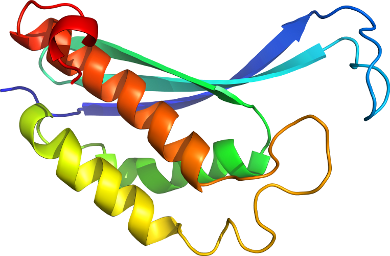
Domain d3pwia1: 3pwi A:7-137 [233288]
Other proteins in same PDB: d3pwia2, d3pwib2
automated match to d1ec7a2
complexed with glr, gol, mg; mutant
Details for d3pwia1
PDB Entry: 3pwi (more details), 2.23 Å
PDB Description: Crystal structure of the mutant P34A of D-Glucarate dehydratase from Escherichia coli complexed with product 5-keto-4-deoxy-D-Glucarate
PDB Compounds: (A:) glucarate dehydrataseSCOPe Domain Sequences for d3pwia1:
Sequence; same for both SEQRES and ATOM records: (download)
>d3pwia1 d.54.1.0 (A:7-137) automated matches {Escherichia coli [TaxId: 444449]}
tpvvtemqvipvaghdsmlmnlsgahaafftrniviikdnsghtgvgeipggekirktle
daiplvvgktlgeyknvltlvrntfadrdaggrglqtfdlrttihvvtgieaamldllgq
hlgvnvasllg
SCOPe Domain Coordinates for d3pwia1:
Click to download the PDB-style file with coordinates for d3pwia1.
(The format of our PDB-style files is described here.)
(The format of our PDB-style files is described here.)
Timeline for d3pwia1:
- d3pwia1 first appeared in SCOPe 2.03
- d3pwia1 appears in SCOPe 2.06
- d3pwia1 appears in the current release, SCOPe 2.08
 View in 3D


