Lineage for d1ek1b1 (1ek1 B:4-225)
- Root: SCOP 1.75

Class c: Alpha and beta proteins (a/b) [51349] (147 folds) 
Fold c.108: HAD-like [56783] (1 superfamily)
3 layers: a/b/a; parallel beta-sheet of 6 strands, order 321456Superfamily c.108.1: HAD-like [56784] (25 families) 
usually contains an insertion (sub)domain after strand 1
Family c.108.1.2: YihX-like [56789] (2 proteins)
the insertion subdomain is a 4-helical bundle
Protein Epoxide hydrolase, N-terminal domain [56790] (2 species)
has a lipid phosphatase activity
Species Mouse (Mus musculus) [TaxId:10090] [56791] (4 PDB entries) 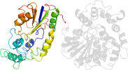
Domain d1ek1b1: 1ek1 B:4-225 [43336]
Other proteins in same PDB: d1ek1a2, d1ek1b2
complexed with ciu
Details for d1ek1b1
PDB Entry: 1ek1 (more details), 3.1 Å
PDB Description: crystal structure of murine soluble epoxide hydrolase complexed with ciu inhibitor
PDB Compounds: (B:) epoxide hydrolaseSCOP Domain Sequences for d1ek1b1:
Sequence; same for both SEQRES and ATOM records: (download)
>d1ek1b1 c.108.1.2 (B:4-225) Epoxide hydrolase, N-terminal domain {Mouse (Mus musculus) [TaxId: 10090]}
rvaafdldgvlalpsiagafrrseealalprdfllgayqtefpegpteqlmkgkitfsqw
vplmdesyrksskacganlpenfsisqifsqamaarsinrpmlqaaialkkkgfttcivt
nnwlddgdkrdslaqmmcelsqhfdfliescqvgmikpepqiynflldtlkakpnevvfl
ddfgsnlkpardmgmvtilvhntasalrelekvtgtqfpeap
SCOP Domain Coordinates for d1ek1b1:
Click to download the PDB-style file with coordinates for d1ek1b1.
(The format of our PDB-style files is described here.)
(The format of our PDB-style files is described here.)
Timeline for d1ek1b1:
- d1ek1b1 first appeared (with stable ids) in SCOP 1.55
- d1ek1b1 appears in SCOP 1.73
- d1ek1b1 appears in SCOPe 2.01
- d1ek1b1 appears in the current release, SCOPe 2.08

 View in 3D


