Lineage for d3usyb1 (3usy B:116-335)
- Root: SCOPe 2.07

Class a: All alpha proteins [46456] (289 folds) 
Fold a.118: alpha-alpha superhelix [48370] (28 superfamilies)
multihelical; 2 (curved) layers: alpha/alpha; right-handed superhelix
Superfamily a.118.14: FliG [48029] (2 families) 
fragmented superhelix; consist of 3/4-helical motifs and connecting helices
Family a.118.14.0: automated matches [191676] (1 protein)
not a true familyProtein automated matches [191304] (1 species)
not a true proteinSpecies Helicobacter pylori [TaxId:210] [190009] (4 PDB entries) 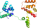
Domain d3usyb1: 3usy B:116-335 [186404]
Other proteins in same PDB: d3usya2, d3usyb2
automated match to d1lkvx_
Details for d3usyb1
PDB Entry: 3usy (more details), 2.71 Å
PDB Description: Crystal structure of Flig (residue 116-343) from H. Pylori
PDB Compounds: (B:) flagellar motor switch proteinSCOPe Domain Sequences for d3usyb1:
Sequence; same for both SEQRES and ATOM records: (download)
>d3usyb1 a.118.14.0 (B:116-335) automated matches {Helicobacter pylori [TaxId: 210]}
qknfaylgkikpqqladfiinehpqtialilahmeapnaaetlsyfpdemkaeisirman
lgeispqvvkrvstvlenklesltsykievgglravaeifnrlgqksakttlariesvdn
klagaikemmftfedivkldnfaireilkvadkkdlslalktstkdltdkflnnmssraa
eqfveemqylgavkikdvdvaqrkiieivqslqekgviqt
SCOPe Domain Coordinates for d3usyb1:
Click to download the PDB-style file with coordinates for d3usyb1.
(The format of our PDB-style files is described here.)
(The format of our PDB-style files is described here.)
Timeline for d3usyb1:
- d3usyb1 first appeared in SCOPe 2.01, called d3usyb_
- d3usyb1 appears in SCOPe 2.06
- d3usyb1 appears in the current release, SCOPe 2.08

 View in 3D


