Lineage for d1ogvl_ (1ogv L:)
- Root: SCOP 1.67

Class f: Membrane and cell surface proteins and peptides [56835] (42 folds) 
Fold f.26: Bacterial photosystem II reaction centre, L and M subunits [81484] (1 superfamily)
five transmembrane helices forming a sheet-like structureSuperfamily f.26.1: Bacterial photosystem II reaction centre, L and M subunits [81483] (1 family) 
Family f.26.1.1: Bacterial photosystem II reaction centre, L and M subunits [81482] (2 proteins)
L and M are probably related to each otherProtein L (light) subunit [81477] (3 species) Species Rhodobacter sphaeroides [TaxId:1063] [81475] (38 PDB entries) 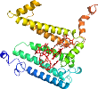
Domain d1ogvl_: 1ogv L: [92950]
Other proteins in same PDB: d1ogvh1, d1ogvh2, d1ogvm_
complexed with bcl, bph, cdl, cl, fe2, u10
Details for d1ogvl_
PDB Entry: 1ogv (more details), 2.35 Å
PDB Description: lipidic cubic phase crystal structure of the photosynthetic reaction centre from rhodobacter sphaeroides
SCOP Domain Sequences for d1ogvl_:
Sequence; same for both SEQRES and ATOM records: (download)
>d1ogvl_ f.26.1.1 (L:) L (light) subunit {Rhodobacter sphaeroides}
allsferkyrvpggtlvggnlfdfwvgpfyvgffgvatfffaalgiiliawsavlqgtwn
pqlisvyppaleyglggaplakgglwqiiticatgafvswalreveicrklgigyhipfa
fafailayltlvlfrpvmmgawgyafpygiwthldwvsntgytygnfhynpahmiaisff
ftnalalalhgalvlsaanpekgkemrtpdhedtffrdlvgysigtlgihrlglllslsa
vffsalcmiitgtiwfdqwvdwwqwwvklpwwanipgging
SCOP Domain Coordinates for d1ogvl_:
Click to download the PDB-style file with coordinates for d1ogvl_.
(The format of our PDB-style files is described here.)
(The format of our PDB-style files is described here.)
Timeline for d1ogvl_:
- d1ogvl_ is new in SCOP 1.67
- d1ogvl_ appears in SCOP 1.69
- d1ogvl_ appears in the current release, SCOPe 2.08

 View in 3D


