Lineage for d1hysa1 (1hys A:430-553)
- Root: SCOP 1.67

Class c: Alpha and beta proteins (a/b) [51349] (130 folds) 
Fold c.55: Ribonuclease H-like motif [53066] (7 superfamilies)
3 layers: a/b/a; mixed beta-sheet of 5 strands, order 32145; strand 2 is antiparallel to the restSuperfamily c.55.3: Ribonuclease H-like [53098] (9 families) 
consists of one domain of this fold
Family c.55.3.1: Ribonuclease H [53099] (3 proteins) 
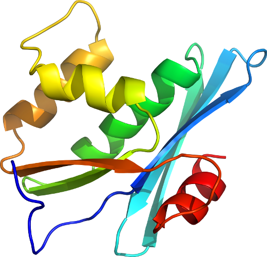
Protein HIV RNase H (Domain of reverse transcriptase) [53105] (2 species) Species Human immunodeficiency virus type 1 [TaxId:11676] [53106] (73 PDB entries) 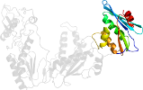
Domain d1hysa1: 1hys A:430-553 [61417]
Other proteins in same PDB: d1hysa2, d1hysb_, d1hysc1, d1hysc2, d1hysd1, d1hysd2
mutant
Details for d1hysa1
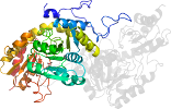
PDB Entry: 1hys (more details), 3 Å
PDB Description: crystal structure of hiv-1 reverse transcriptase in complex with a polypurine tract rna:dna
SCOP Domain Sequences for d1hysa1:
Sequence; same for both SEQRES and ATOM records: (download)
>d1hysa1 c.55.3.1 (A:430-553) HIV RNase H (Domain of reverse transcriptase) {Human immunodeficiency virus type 1}
ekepivgaetfyvdgaanretklgkagyvtnkgrqkvvpltnttnqktelqaiylalqds
glevnivtdsqyalgiiqaqpdkseselvnqiieqlikkekvylawvpahkgiggneqvd
klvs
SCOP Domain Coordinates for d1hysa1:
Click to download the PDB-style file with coordinates for d1hysa1.
(The format of our PDB-style files is described here.)
(The format of our PDB-style files is described here.)
Timeline for d1hysa1:
- d1hysa1 first appeared in SCOP 1.57
- d1hysa1 appears in SCOP 1.65
- d1hysa1 appears in SCOP 1.69
- d1hysa1 appears in the current release, SCOPe 2.08
 View in 3D