Lineage for d1hsad2 (1hsa D:1-181)
- Root: SCOP 1.67

Class d: Alpha and beta proteins (a+b) [53931] (260 folds) 
Fold d.19: MHC antigen-recognition domain [54451] (1 superfamily)
dimericSuperfamily d.19.1: MHC antigen-recognition domain [54452] (1 family) 
Family d.19.1.1: MHC antigen-recognition domain [54453] (11 proteins) Protein Class I MHC, alpha-1 and alpha-2 domains [54468] (20 species) Species Human (Homo sapiens), HLA-B27 [TaxId:9606] [54471] (6 PDB entries) 
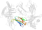
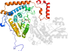
Domain d1hsad2: 1hsa D:1-181 [38256]
Other proteins in same PDB: d1hsaa1, d1hsab_, d1hsad1, d1hsae_
Details for d1hsad2
PDB Entry: 1hsa (more details), 2.1 Å
PDB Description: the three-dimensional structure of hla-b27 at 2.1 angstroms resolution suggests a general mechanism for tight peptide binding to mhc
SCOP Domain Sequences for d1hsad2:
Sequence; same for both SEQRES and ATOM records: (download)
>d1hsad2 d.19.1.1 (D:1-181) Class I MHC, alpha-1 and alpha-2 domains {Human (Homo sapiens), HLA-B27}
gshsmryfhtsvsrpgrgeprfitvgyvddtlfvrfdsdaaspreeprapwieqegpeyw
dretqickakaqtdredlrtllryynqseagshtlqnmygcdvgpdgrllrgyhqdaydg
kdyialnedlsswtaadtaaqitqrkweaarvaeqlraylegecvewlrrylengketlq
r
SCOP Domain Coordinates for d1hsad2:
Click to download the PDB-style file with coordinates for d1hsad2.
(The format of our PDB-style files is described here.)
(The format of our PDB-style files is described here.)
Timeline for d1hsad2:
- d1hsad2 first appeared (with stable ids) in SCOP 1.55
- d1hsad2 appears in SCOP 1.65
- d1hsad2 appears in SCOP 1.69
- d1hsad2 appears in the current release, SCOPe 2.08
 View in 3D View in 3DDomains from other chains: (mouse over for more information) d1hsaa1, d1hsaa2, d1hsab_, d1hsae_ |