Lineage for d2h1pl2 (2h1p L:114-219)
- Root: SCOP 1.71

Class b: All beta proteins [48724] (149 folds) 
Fold b.1: Immunoglobulin-like beta-sandwich [48725] (25 superfamilies)
sandwich; 7 strands in 2 sheets; greek-key
some members of the fold have additional strandsSuperfamily b.1.1: Immunoglobulin [48726] (4 families) 
Family b.1.1.2: C1 set domains (antibody constant domain-like) [48942] (23 proteins) 
Protein Immunoglobulin light chain kappa constant domain, CL-kappa [88566] (3 species) Species Mouse (Mus musculus) [TaxId:10090] [88567] (284 PDB entries) 
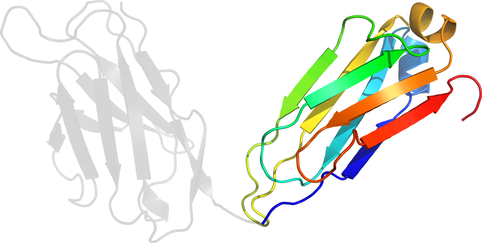
Domain d2h1pl2: 2h1p L:114-219 [21206]
Other proteins in same PDB: d2h1ph1, d2h1ph2, d2h1pl1
part of polysaccharide binding antibody 2H1
Details for d2h1pl2
PDB Entry: 2h1p (more details), 2.4 Å
PDB Description: the three-dimensional structures of a polysaccharide binding antibody to cryptococcus neoformans and its complex with a peptide from a phage display library: implications for the identification of peptide mimotopes
SCOP Domain Sequences for d2h1pl2:
Sequence; same for both SEQRES and ATOM records: (download)
>d2h1pl2 b.1.1.2 (L:114-219) Immunoglobulin light chain kappa constant domain, CL-kappa {Mouse (Mus musculus)}
adaaptvsifppsseqltsggasvvcflnnfypkdinvkwkidgserqngvlnswtdeds
kdstysmsstltltkdeyerhnsytceathktstspivksfnrnec
SCOP Domain Coordinates for d2h1pl2:
Click to download the PDB-style file with coordinates for d2h1pl2.
(The format of our PDB-style files is described here.)
(The format of our PDB-style files is described here.)
Timeline for d2h1pl2:
- d2h1pl2 first appeared (with stable ids) in SCOP 1.55
- d2h1pl2 appears in SCOP 1.69
- d2h1pl2 appears in SCOP 1.73
- d2h1pl2 appears in the current release, SCOPe 2.08

 View in 3D