Lineage for d2h1pl1 (2h1p L:1-113)
- Root: SCOP 1.61

Class b: All beta proteins [48724] (111 folds) 
Fold b.1: Immunoglobulin-like beta-sandwich [48725] (17 superfamilies) 
Superfamily b.1.1: Immunoglobulin [48726] (6 families) 
Family b.1.1.1: V set domains (antibody variable domain-like) [48727] (14 proteins) 
Protein Immunoglobulin (variable domains of L and H chains) [48749] (222 species) 
Species Fab 2H1 (mouse), kappa L chain [48837] (1 PDB entry) 
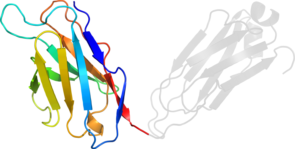
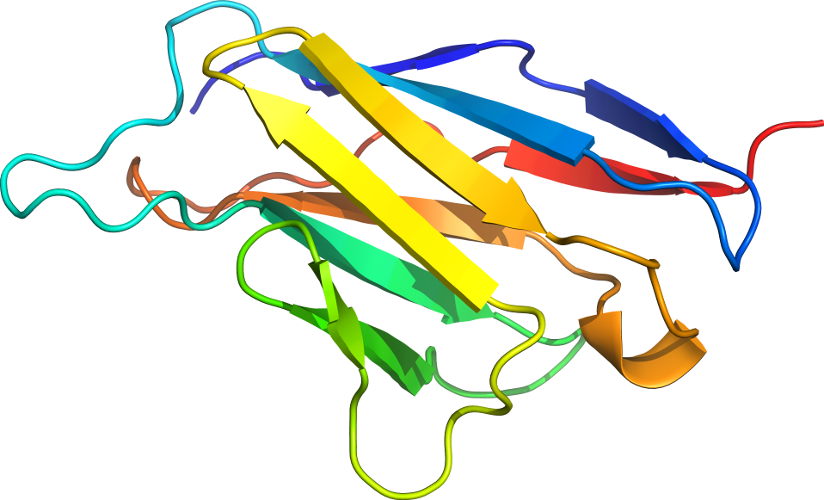
Domain d2h1pl1: 2h1p L:1-113 [20190]
Other proteins in same PDB: d2h1ph2, d2h1pl2
Details for d2h1pl1
PDB Entry: 2h1p (more details), 2.4 Å
PDB Description: the three-dimensional structures of a polysaccharide binding antibody to cryptococcus neoformans and its complex with a peptide from a phage display library: implications for the identification of peptide mimotopes
SCOP Domain Sequences for d2h1pl1:
Sequence; same for both SEQRES and ATOM records: (download)
>d2h1pl1 b.1.1.1 (L:1-113) Immunoglobulin (variable domains of L and H chains) {Fab 2H1 (mouse), kappa L chain}
dvvmtqtplslpvslgdpasiscrssqslvhsngntylhwylqkpgqspklliykvsnrf
sgvpdkfsgsgsgtdftlkisrveaedqgvyfcsqsthvpwtfgggtkleikr
SCOP Domain Coordinates for d2h1pl1:
Click to download the PDB-style file with coordinates for d2h1pl1.
(The format of our PDB-style files is described here.)
(The format of our PDB-style files is described here.)
Timeline for d2h1pl1:
- d2h1pl1 first appeared (with stable ids) in SCOP 1.55
- d2h1pl1 appears in SCOP 1.59
- d2h1pl1 appears in SCOP 1.63
- d2h1pl1 appears in the current release, SCOPe 2.08
 View in 3D