Lineage for d2qp0i1 (2qp0 I:3-129)
- Root: SCOPe 2.03

Class d: Alpha and beta proteins (a+b) [53931] (376 folds) 
Fold d.14: Ribosomal protein S5 domain 2-like [54210] (1 superfamily)
core: beta(3)-alpha-beta-alpha; 2 layers: alpha/beta; left-handed crossoverSuperfamily d.14.1: Ribosomal protein S5 domain 2-like [54211] (13 families) 

Family d.14.1.1: Translational machinery components [54212] (4 proteins) 
Protein Ribosomal protein S9 [54218] (2 species) 
Species Escherichia coli [TaxId:562] [159907] (26 PDB entries)
Uniprot P0A7X3 3-129
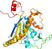
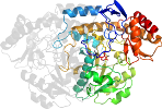
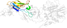
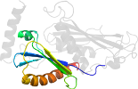
Domain d2qp0i1: 2qp0 I:3-129 [151152]
Other proteins in same PDB: d2qp0b1, d2qp0c1, d2qp0c2, d2qp0d1, d2qp0e1, d2qp0e2, d2qp0f1, d2qp0g1, d2qp0h1, d2qp0j1, d2qp0k1, d2qp0l1, d2qp0m1, d2qp0n1, d2qp0p1, d2qp0q1, d2qp0r1, d2qp0s1, d2qp0t1, d2qp0u1
automatically matched to 2AVY I:3-129
protein/RNA complex; complexed with mg, nmy, scm
Details for d2qp0i1
PDB Entry: 2qp0 (more details), 3.5 Å
PDB Description: Crystal structure of the bacterial ribosome from Escherichia coli in complex with spectinomycin and neomycin. This file contains the 30S subunit of the second 70S ribosome, with spectinomycin and neomycin bound. The entire crystal structure contains two 70S ribosomes.
PDB Compounds: (I:) 30S ribosomal protein S9SCOPe Domain Sequences for d2qp0i1:
Sequence; same for both SEQRES and ATOM records: (download)
>d2qp0i1 d.14.1.1 (I:3-129) Ribosomal protein S9 {Escherichia coli [TaxId: 562]}
nqyygtgrrkssaarvfikpgngkivinqrsleqyfgretarmvvrqplelvdmvekldl
yitvkgggisgqagairhgitralmeydeslrselrkagfvtrdarqverkkvglrkarr
rpqfskr
SCOPe Domain Coordinates for d2qp0i1:
Click to download the PDB-style file with coordinates for d2qp0i1.
(The format of our PDB-style files is described here.)
(The format of our PDB-style files is described here.)
Timeline for d2qp0i1:
- d2qp0i1 first appeared in SCOP 1.75
- d2qp0i1 appears in SCOPe 2.02
- d2qp0i1 appears in SCOPe 2.04
- d2qp0i1 appears in the current release, SCOPe 2.08

 View in 3D