Lineage for d1vs6f1 (1vs6 F:1-178)
- Root: SCOPe 2.03

Class d: Alpha and beta proteins (a+b) [53931] (376 folds) 
Fold d.77: RL5-like [55281] (1 superfamily)
beta-alpha-beta(2)-alpha-beta(3)-alpha; 2 layers, alpha/beta; antiparallel beta-sheet: order 231654Superfamily d.77.1: RL5-like [55282] (3 families) 
Family d.77.1.1: Ribosomal protein L5 [55283] (1 protein) Protein Ribosomal protein L5 [55284] (5 species)
synonym: 50S ribosomal protein L5p, HMAL5, HL13
Species Escherichia coli [TaxId:562] [160488] (29 PDB entries)
Uniprot P62399 1-178
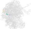
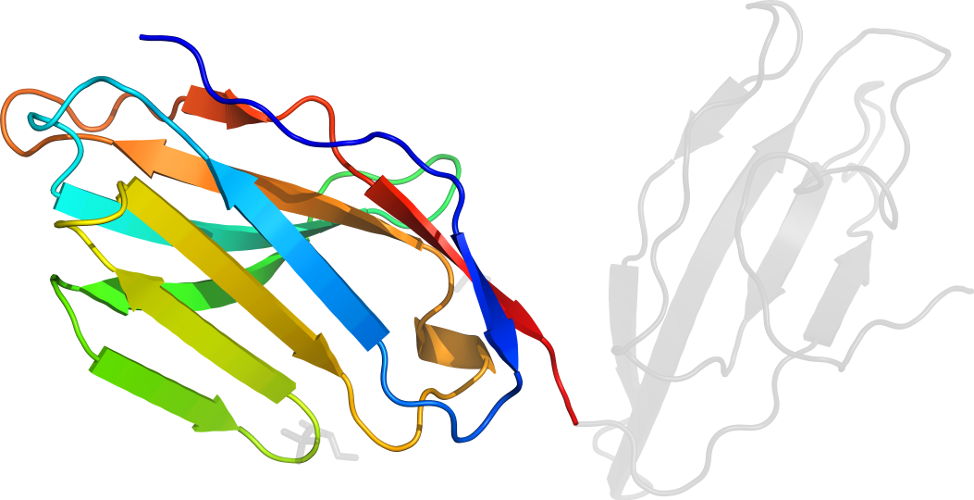
Domain d1vs6f1: 1vs6 F:1-178 [144460]
Other proteins in same PDB: d1vs601, d1vs611, d1vs621, d1vs631, d1vs641, d1vs6c1, d1vs6c2, d1vs6d1, d1vs6e1, d1vs6g1, d1vs6g2, d1vs6h1, d1vs6h2, d1vs6i1, d1vs6i2, d1vs6j1, d1vs6k1, d1vs6l1, d1vs6m1, d1vs6n1, d1vs6o1, d1vs6p1, d1vs6q1, d1vs6r1, d1vs6s1, d1vs6t1, d1vs6u1, d1vs6v1, d1vs6w1, d1vs6x1, d1vs6y1, d1vs6z1
automatically matched to 2AW4 F:1-178
complexed with mg
Details for d1vs6f1
PDB Entry: 1vs6 (more details), 3.46 Å
PDB Description: Crystal structure of the bacterial ribosome from escherichia coli in complex with the antibiotic kasugamyin at 3.5A resolution. this file contains the 50s subunit of one 70s ribosome. the entire crystal structure contains two 70s ribosomes and is described in remark 400.
PDB Compounds: (F:) 50S ribosomal protein L5SCOPe Domain Sequences for d1vs6f1:
Sequence; same for both SEQRES and ATOM records: (download)
>d1vs6f1 d.77.1.1 (F:1-178) Ribosomal protein L5 {Escherichia coli [TaxId: 562]}
aklhdyykdevvkklmtefnynsvmqvprvekitlnmgvgeaiadkklldnaaadlaais
gqkplitkarksvagfkirqgypigckvtlrgermwefferlitiavprirdfrglsaks
fdgrgnysmgvreqiifpeidydkvdrvrgldititttaksdeegrallaafdfpfrk
SCOPe Domain Coordinates for d1vs6f1:
Click to download the PDB-style file with coordinates for d1vs6f1.
(The format of our PDB-style files is described here.)
(The format of our PDB-style files is described here.)
Timeline for d1vs6f1:
- d1vs6f1 first appeared in SCOP 1.75
- d1vs6f1 appears in SCOPe 2.02
- d1vs6f1 appears in SCOPe 2.04
- d1vs6f1 appears in the current release, SCOPe 2.08
 View in 3D View in 3DDomains from other chains: (mouse over for more information) d1vs601, d1vs611, d1vs621, d1vs631, d1vs641, d1vs6c1, d1vs6c2, d1vs6d1, d1vs6e1, d1vs6g1, d1vs6g2, d1vs6h1, d1vs6h2, d1vs6i1, d1vs6i2, d1vs6j1, d1vs6k1, d1vs6l1, d1vs6m1, d1vs6n1, d1vs6o1, d1vs6p1, d1vs6q1, d1vs6r1, d1vs6s1, d1vs6t1, d1vs6u1, d1vs6v1, d1vs6w1, d1vs6x1, d1vs6y1, d1vs6z1 |