Lineage for d1vs5c2 (1vs5 C:106-206)
- Root: SCOP 1.75

Class d: Alpha and beta proteins (a+b) [53931] (376 folds) 
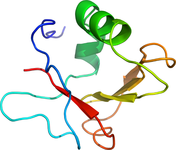
Fold d.53: Ribosomal protein S3 C-terminal domain [54820] (1 superfamily)
alpha(2)-beta(4); 2 layers: alpha/beta; antiparallel beta-sheet: order 2143Superfamily d.53.1: Ribosomal protein S3 C-terminal domain [54821] (1 family) 
Family d.53.1.1: Ribosomal protein S3 C-terminal domain [54822] (1 protein) Protein Ribosomal protein S3 C-terminal domain [54823] (2 species) 
Species Escherichia coli [TaxId:562] [160263] (24 PDB entries)
Uniprot P0A7V3 106-206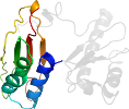
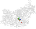
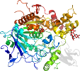
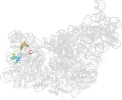
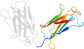
Domain d1vs5c2: 1vs5 C:106-206 [144437]
Other proteins in same PDB: d1vs5b1, d1vs5c1, d1vs5d1, d1vs5e1, d1vs5e2, d1vs5f1, d1vs5g1, d1vs5h1, d1vs5i1, d1vs5j1, d1vs5k1, d1vs5l1, d1vs5m1, d1vs5n1, d1vs5o1, d1vs5p1, d1vs5q1, d1vs5r1, d1vs5s1, d1vs5t1, d1vs5u1
automatically matched to 2AVY C:106-206
complexed with ksg, mg
Details for d1vs5c2
PDB Entry: 1vs5 (more details), 3.46 Å
PDB Description: Crystal structure of the bacterial ribosome from escherichia coli in complex with the antibiotic kasugamyin at 3.5A resolution. this file contains the 30s subunit of one 70s ribosome. the entire crystal structure contains two 70s ribosomes and is described in remark 400.
PDB Compounds: (C:) 30S ribosomal protein S3SCOP Domain Sequences for d1vs5c2:
Sequence; same for both SEQRES and ATOM records: (download)
>d1vs5c2 d.53.1.1 (C:106-206) Ribosomal protein S3 C-terminal domain {Escherichia coli [TaxId: 562]}
rkpeldaklvadsitsqlerrvmfrramkravqnamrlgakgikvevsgrlggaeiarte
wyregrvplhtlradidyntseahttygvigvkvwifkgei
SCOP Domain Coordinates for d1vs5c2:
Click to download the PDB-style file with coordinates for d1vs5c2.
(The format of our PDB-style files is described here.)
(The format of our PDB-style files is described here.)
Timeline for d1vs5c2:
- d1vs5c2 is new in SCOP 1.75
- d1vs5c2 appears in SCOPe 2.01
- d1vs5c2 appears in the current release, SCOPe 2.08

 View in 3D