Lineage for d1vldr1 (1vld R:196-274)
- Root: SCOP 1.69

Class b: All beta proteins [48724] (144 folds) 
Fold b.3: Prealbumin-like [49451] (6 superfamilies)
sandwich; 7 strands in 2 sheets, greek-key
variations: some members have additional 1-2 strands to common fold
Superfamily b.3.5: Cna protein B-type domain (Pfam 05738) [49478] (1 family) 
Family b.3.5.1: Cna protein B-type domain (Pfam 05738) [49479] (2 proteins) Protein Transhydroxylase beta subunit, BthL, C-terminal domain [110094] (1 species)
the penultimate strand is in the other beta-sheet than in the Cna repeatsSpecies Pelobacter acidigallici [TaxId:35816] [110095] (6 PDB entries) 
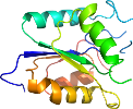
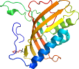
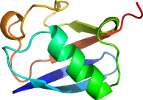
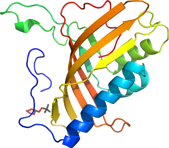
Domain d1vldr1: 1vld R:196-274 [108754]
Other proteins in same PDB: d1vldm1, d1vldm2, d1vldn2, d1vldo1, d1vldo2, d1vldp2, d1vldq1, d1vldq2, d1vldr2, d1vlds1, d1vlds2, d1vldt2, d1vldu1, d1vldu2, d1vldv2, d1vldw1, d1vldw2, d1vldx2
Details for d1vldr1
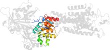
PDB Entry: 1vld (more details), 2.35 Å
PDB Description: Crystal Structure of Pyrogallol-Phloroglucinol Transhydroxylase from Pelobacter acidigallici
SCOP Domain Sequences for d1vldr1:
Sequence; same for both SEQRES and ATOM records: (download)
>d1vldr1 b.3.5.1 (R:196-274) Transhydroxylase beta subunit, BthL, C-terminal domain {Pelobacter acidigallici}
knyvtagilvqgdcfegakvvlksggkevasaetnffgefkfdaldngeytveidadgks
ysdtvviddksvdlgfikl
SCOP Domain Coordinates for d1vldr1:
Click to download the PDB-style file with coordinates for d1vldr1.
(The format of our PDB-style files is described here.)
(The format of our PDB-style files is described here.)
Timeline for d1vldr1:
- d1vldr1 is new in SCOP 1.69
- d1vldr1 appears in SCOP 1.71
- d1vldr1 appears in the current release, SCOPe 2.08

 View in 3D