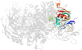
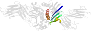
Lineage for d1r5wd2 (1r5w D:31-120)
- Root: SCOPe 2.02

Class d: Alpha and beta proteins (a+b) [53931] (376 folds) 
Fold d.19: MHC antigen-recognition domain [54451] (1 superfamily)
dimericSuperfamily d.19.1: MHC antigen-recognition domain [54452] (1 family) 
Family d.19.1.1: MHC antigen-recognition domain [54453] (13 proteins) 
Protein Class II MHC beta chain, N-terminal domain [88819] (15 species) 
Species Mouse (Mus musculus), I-EK [TaxId:10090] [88827] (9 PDB entries) 
Domain d1r5wd2: 1r5w D:31-120 [97131]
Other proteins in same PDB: d1r5wa1, d1r5wa2, d1r5wb1, d1r5wc1, d1r5wc2, d1r5wd1
Details for d1r5wd2
PDB Entry: 1r5w (more details), 2.9 Å
PDB Description: evidence that structural rearrangements and/or flexibility during tcr binding can contribute to t-cell activation
PDB Compounds: (D:) MHC H2-IE-betaSCOPe Domain Sequences for d1r5wd2:
Sequence; same for both SEQRES and ATOM records: (download)
>d1r5wd2 d.19.1.1 (D:31-120) Class II MHC beta chain, N-terminal domain {Mouse (Mus musculus), I-EK [TaxId: 10090]}
apwfleysksechfyngtqrvrllvryfynleenlrfdsdvgefravtelgrpdaenwns
qpefleqkraevdtvcrhnyeifdnflvpr
SCOPe Domain Coordinates for d1r5wd2:
Click to download the PDB-style file with coordinates for d1r5wd2.
(The format of our PDB-style files is described here.)
(The format of our PDB-style files is described here.)
Timeline for d1r5wd2:
- d1r5wd2 first appeared in SCOP 1.67
- d1r5wd2 appears in SCOPe 2.01
- d1r5wd2 appears in SCOPe 2.03
- d1r5wd2 appears in the current release, SCOPe 2.08

 View in 3D