Lineage for d1ogvh2 (1ogv H:11-35)
- Root: SCOPe 2.08

Class f: Membrane and cell surface proteins and peptides [56835] (69 folds) 
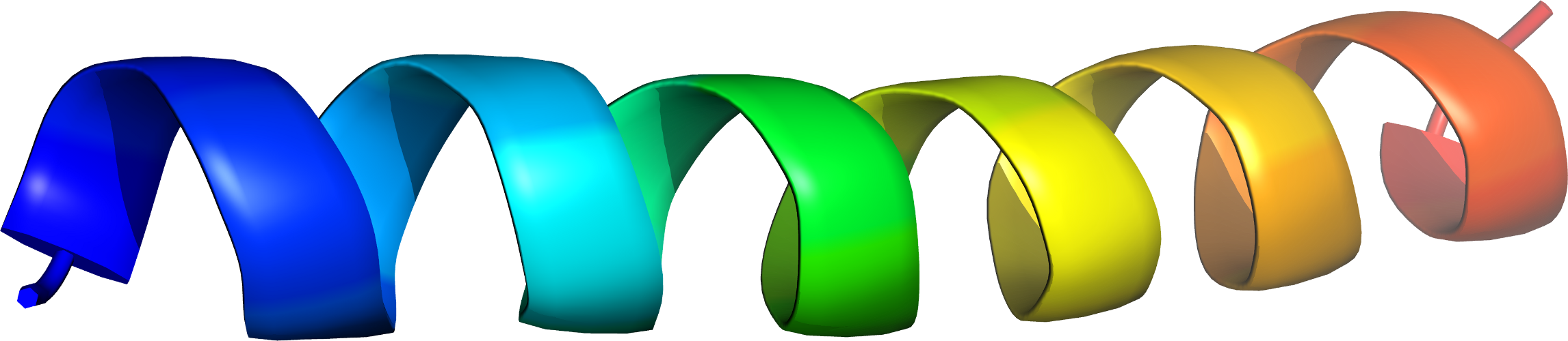
Fold f.23: Single transmembrane helix [81407] (42 superfamilies)
not a true fold
annotated by the SCOP(e) curators as 'not a true fold'
Superfamily f.23.10: Photosystem II reaction centre subunit H, transmembrane region [81490] (2 families) 
Family f.23.10.1: Photosystem II reaction centre subunit H, transmembrane region [81489] (1 protein) Protein Photosystem II reaction centre subunit H, transmembrane region [81488] (4 species) 
Species Rhodobacter sphaeroides [TaxId:1063] [81486] (88 PDB entries)
Uniprot P11846
Domain d1ogvh2: 1ogv H:11-35 [92949]
Other proteins in same PDB: d1ogvh1, d1ogvl_, d1ogvm_
complexed with bcl, bph, cdl, cl, fe2, u10
Details for d1ogvh2
PDB Entry: 1ogv (more details), 2.35 Å
PDB Description: lipidic cubic phase crystal structure of the photosynthetic reaction centre from rhodobacter sphaeroides
PDB Compounds: (H:) reaction center protein h chainSCOPe Domain Sequences for d1ogvh2:
Sequence; same for both SEQRES and ATOM records: (download)
>d1ogvh2 f.23.10.1 (H:11-35) Photosystem II reaction centre subunit H, transmembrane region {Rhodobacter sphaeroides [TaxId: 1063]}
dlaslaiysfwiflagliyylqten
SCOPe Domain Coordinates for d1ogvh2:
Click to download the PDB-style file with coordinates for d1ogvh2.
(The format of our PDB-style files is described here.)
(The format of our PDB-style files is described here.)
Timeline for d1ogvh2:
- d1ogvh2 first appeared in SCOP 1.67
- d1ogvh2 appears in SCOPe 2.07
 View in 3D


