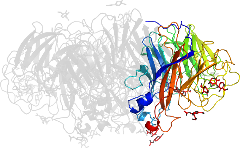
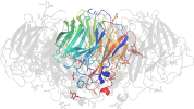
Lineage for d1hm0a1 (1hm0 A:252-447)
- Root: SCOP 1.63

Class b: All beta proteins [48724] (119 folds) 
Fold b.81: Single-stranded left-handed beta-helix [51160] (2 superfamilies)
superhelix with each turn made by 3 strands with short linksSuperfamily b.81.1: Trimeric LpxA-like enzymes [51161] (5 families) 
duplication: the sequence hexapeptide repeats correspond to individual strands
Family b.81.1.4: N-acetylglucosamine 1-phosphate uridyltransferase GlmU, C-terminal domain [51171] (1 protein)
this is a repeat family; one repeat unit is 2oi5 A:321-339 found in domainProtein N-acetylglucosamine 1-phosphate uridyltransferase GlmU, C-terminal domain [51172] (2 species) Species Streptococcus pneumoniae [TaxId:1313] [63852] (5 PDB entries) 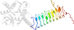
Domain d1hm0a1: 1hm0 A:252-447 [65858]
Other proteins in same PDB: d1hm0a2, d1hm0b2
Details for d1hm0a1
PDB Entry: 1hm0 (more details), 2.3 Å
PDB Description: crystal structure of s.pneumoniae n-acetylglucosamine 1-phosphate uridyltransferase, glmu
SCOP Domain Sequences for d1hm0a1:
Sequence; same for both SEQRES and ATOM records: (download)
>d1hm0a1 b.81.1.4 (A:252-447) N-acetylglucosamine 1-phosphate uridyltransferase GlmU, C-terminal domain {Streptococcus pneumoniae}
vsfvnpeatyididveiapevqieanvilkgqtkigaetvltngtyvvdstigagavitn
smieessvadgvtvgpyahirpnsslgaqvhignfvevkgssigentkaghltyigncev
gsnvnfgagtitvnydgknkyktvigdnvfvgsnstiiapvelgdnslvgagstitkdvp
adaiaigrgrqinkde
SCOP Domain Coordinates for d1hm0a1:
Click to download the PDB-style file with coordinates for d1hm0a1.
(The format of our PDB-style files is described here.)
(The format of our PDB-style files is described here.)
Timeline for d1hm0a1:
- d1hm0a1 first appeared in SCOP 1.59
- d1hm0a1 appears in SCOP 1.61
- d1hm0a1 appears in SCOP 1.65
- d1hm0a1 appears in the current release, SCOPe 2.08

 View in 3D