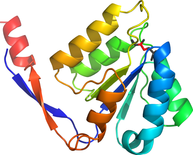
Lineage for d1ik4c_ (1ik4 C:)
- Root: SCOPe 2.08

Class c: Alpha and beta proteins (a/b) [51349] (148 folds) 
Fold c.24: Methylglyoxal synthase-like [52334] (1 superfamily)
3 layers, a/b/a; parallel beta-sheet of 5 strands, order 32145Superfamily c.24.1: Methylglyoxal synthase-like [52335] (4 families) 
contains a common phosphate-binding siteFamily c.24.1.2: Methylglyoxal synthase, MgsA [52339] (2 proteins) 
Protein Methylglyoxal synthase, MgsA [52340] (3 species) 
Species Escherichia coli [TaxId:562] [52341] (5 PDB entries) 
Domain d1ik4c_: 1ik4 C: [62521]
complexed with phosphoglycolohydroxamic acid
complexed with pgh
Details for d1ik4c_
PDB Entry: 1ik4 (more details), 2 Å
PDB Description: X-ray Structure of Methylglyoxal Synthase from E. coli Complexed with Phosphoglycolohydroxamic Acid
PDB Compounds: (C:) methylglyoxal synthaseSCOPe Domain Sequences for d1ik4c_:
Sequence; same for both SEQRES and ATOM records: (download)
>d1ik4c_ c.24.1.2 (C:) Methylglyoxal synthase, MgsA {Escherichia coli [TaxId: 562]}
melttrtlparkhialvahdhckqmlmswverhqplleqhvlyatgttgnlisratgmnv
namlsgpmggdqqvgalisegkidvliffwdplnavphdpdvkallrlatvwnipvatnv
atadfiiqsphfndavdilipdyqryladrlk
SCOPe Domain Coordinates for d1ik4c_:
Click to download the PDB-style file with coordinates for d1ik4c_.
(The format of our PDB-style files is described here.)
(The format of our PDB-style files is described here.)
Timeline for d1ik4c_:
- d1ik4c_ first appeared in SCOP 1.57
- d1ik4c_ appears in SCOPe 2.07

 View in 3D