Lineage for d1pbba2 (1pbb A:174-275)
- Root: SCOP 1.75

Class d: Alpha and beta proteins (a+b) [53931] (376 folds) 
Fold d.16: FAD-linked reductases, C-terminal domain [54372] (1 superfamily)
alpha+beta sandwichSuperfamily d.16.1: FAD-linked reductases, C-terminal domain [54373] (7 families) 
N-terminal domain is beta/beta/alpha common fold
Family d.16.1.2: PHBH-like [54378] (4 proteins) 
Protein p-Hydroxybenzoate hydroxylase (PHBH) [54379] (2 species) 
Species Pseudomonas fluorescens [TaxId:294] [54380] (17 PDB entries) 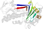
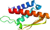
Domain d1pbba2: 1pbb A:174-275 [37879]
Other proteins in same PDB: d1pbba1
complexed with dob, fad; mutant
Details for d1pbba2
PDB Entry: 1pbb (more details), 2.5 Å
PDB Description: crystal structures of wild-type p-hydroxybenzoate hydroxylase complexed with 4-aminobenzoate, 2,4-dihydroxybenzoate and 2-hydroxy- 4-aminobenzoate and of the try222ala mutant, complexed with 2- hydroxy-4-aminobenzoate. evidence for a proton channel and a new binding mode of the flavin ring
PDB Compounds: (A:) p-hydroxybenzoate hydroxylaseSCOP Domain Sequences for d1pbba2:
Sequence; same for both SEQRES and ATOM records: (download)
>d1pbba2 d.16.1.2 (A:174-275) p-Hydroxybenzoate hydroxylase (PHBH) {Pseudomonas fluorescens [TaxId: 294]}
lkvfervypfgwlglladtppvsheliyanhprgfalcsqrsatrsryyvqvpltekved
wsderfwtelkarlpaevaeklvtgpsleksiaplrsfvvep
SCOP Domain Coordinates for d1pbba2:
Click to download the PDB-style file with coordinates for d1pbba2.
(The format of our PDB-style files is described here.)
(The format of our PDB-style files is described here.)
Timeline for d1pbba2:
- d1pbba2 first appeared (with stable ids) in SCOP 1.55, called d1pbb_2
- d1pbba2 appears in SCOP 1.73
- d1pbba2 appears in SCOPe 2.01
- d1pbba2 appears in the current release, SCOPe 2.08

 View in 3D