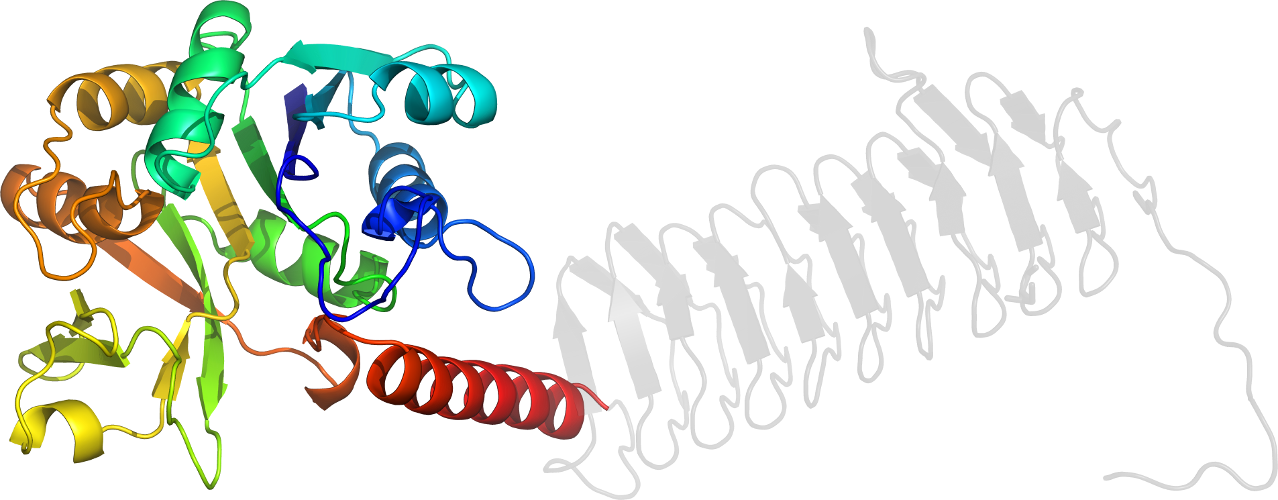
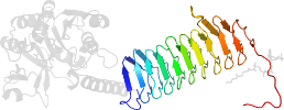
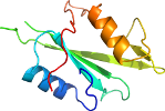
Lineage for d1hv9a2 (1hv9 A:4-251)
- Root: SCOP 1.71

Class c: Alpha and beta proteins (a/b) [51349] (134 folds) 
Fold c.68: Nucleotide-diphospho-sugar transferases [53447] (1 superfamily)
3 layers: a/b/a; mixed beta-sheet of 7 strands, order 3214657; strand 6 is antiparallel to the restSuperfamily c.68.1: Nucleotide-diphospho-sugar transferases [53448] (14 families) 

Family c.68.1.5: UDP-glucose pyrophosphorylase [53461] (2 proteins) 
Protein N-acetylglucosamine 1-phosphate uridyltransferase GlmU, N-terminal domain [53462] (2 species) 
Species Escherichia coli [TaxId:562] [53463] (3 PDB entries) 
Domain d1hv9a2: 1hv9 A:4-251 [34518]
Other proteins in same PDB: d1hv9a1, d1hv9b1
Details for d1hv9a2
PDB Entry: 1hv9 (more details), 2.1 Å
PDB Description: structure of e. coli glmu: analysis of pyrophosphorylase and acetyltransferase active sites
SCOP Domain Sequences for d1hv9a2:
Sequence; same for both SEQRES and ATOM records: (download)
>d1hv9a2 c.68.1.5 (A:4-251) N-acetylglucosamine 1-phosphate uridyltransferase GlmU, N-terminal domain {Escherichia coli}
namsvvilaagkgtrmysdlpkvlhtlagkamvqhvidaanelgaahvhlvyghggdllk
qalkddnlnwvlqaeqlgtghamqqaapffaddedilmlygdvplisvetlqrlrdakpq
ggiglltvklddptgygritrengkvtgivehkdatdeqrqiqeintgiliangadmkrw
lakltnnnaqgeyyitdiialayqegreivavhpqrlsevegvnnrlqlsrlervyqseq
aeklllag
SCOP Domain Coordinates for d1hv9a2:
Click to download the PDB-style file with coordinates for d1hv9a2.
(The format of our PDB-style files is described here.)
(The format of our PDB-style files is described here.)
Timeline for d1hv9a2:
- d1hv9a2 first appeared (with stable ids) in SCOP 1.55
- d1hv9a2 appears in SCOP 1.69
- d1hv9a2 appears in SCOP 1.73
- d1hv9a2 appears in the current release, SCOPe 2.08

 View in 3D