Lineage for d1pbd_1 (1pbd 1-173,276-391)
- Root: SCOP 1.55

Class c: Alpha and beta proteins (a/b) [51349] (97 folds) 
Fold c.3: FAD/NAD(P)-binding domain [51904] (1 superfamily) Superfamily c.3.1: FAD/NAD(P)-binding domain [51905] (5 families) 

Family c.3.1.2: FAD-linked reductases, N-terminal domain [51913] (7 proteins) 
Protein p-Hydroxybenzoate hydroxylase (PHBH) [51917] (2 species) Species Pseudomonas fluorescens [TaxId:294] [51918] (17 PDB entries) 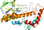
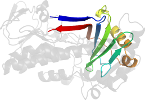
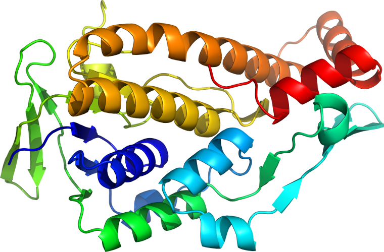
Domain d1pbd_1: 1pbd 1-173,276-391 [30342]
Other proteins in same PDB: d1pbd_2
Details for d1pbd_1
PDB Entry: 1pbd (more details), 2.3 Å
PDB Description: crystal structures of wild-type p-hydroxybenzoate hydroxylase complexed with 4-aminobenzoate, 2,4-dihydroxybenzoate and 2-hydroxy- 4-aminobenzoate and of the try222ala mutant, complexed with 2- hydroxy-4-aminobenzoate. evidence for a proton channel and a new binding mode of the flavin ring
SCOP Domain Sequences for d1pbd_1:
Sequence; same for both SEQRES and ATOM records: (download)
>d1pbd_1 c.3.1.2 (1-173,276-391) p-Hydroxybenzoate hydroxylase (PHBH) {Pseudomonas fluorescens}
mktqvaiigagpsglllgqllhkagidnvilerqtpdyvlgriragvleqgmvdllreag
vdrrmardglvhegveiafagqrrridlkrlsggktvtvygqtevtrdlmeareasgatt
vyqaaevrlhdlqgerpyvtferdgerlrldcdyiagcdgfhgisrqsipaerXmqhgrl
flagdaahivpptgakglnlaasdvstlyrlllkayregrgellerysaiclrriwkaer
fswwmtsvlhrfpdtdafsqriqqteleyylgseaglatiaenyvglpye
SCOP Domain Coordinates for d1pbd_1:
Click to download the PDB-style file with coordinates for d1pbd_1.
(The format of our PDB-style files is described here.)
(The format of our PDB-style files is described here.)
Timeline for d1pbd_1:
- d1pbd_1 appears in SCOP 1.57
- d1pbd_1 appears in the current release, SCOPe 2.08, called d1pbda1
 View in 3D