Lineage for d1fwyb1 (1fwy B:252-326)
- Root: SCOPe 2.01

Class b: All beta proteins [48724] (174 folds) 
Fold b.81: Single-stranded left-handed beta-helix [51160] (4 superfamilies)
superhelix turns are made of parallel beta-strands and (short) turnsSuperfamily b.81.1: Trimeric LpxA-like enzymes [51161] (9 families) 
superhelical turns are made of three short strands; duplication: the sequence hexapeptide repeats correspond to individual strands
Family b.81.1.4: GlmU C-terminal domain-like [51171] (3 proteins)
this is a repeat family; one repeat unit is 2oi5 A:321-339 found in domain
Protein N-acetylglucosamine 1-phosphate uridyltransferase GlmU, C-terminal domain [51172] (2 species) 
Species Escherichia coli [TaxId:562] [51173] (6 PDB entries) 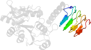
Domain d1fwyb1: 1fwy B:252-326 [28065]
Other proteins in same PDB: d1fwya2, d1fwyb2
truncated form after R331
complexed with edo, so4, ud1
Details for d1fwyb1
PDB Entry: 1fwy (more details), 2.3 Å
PDB Description: crystal structure of n-acetylglucosamine 1-phosphate uridyltransferase bound to udp-glcnac
PDB Compounds: (B:) udp-n-acetylglucosamine pyrophosphorylaseSCOPe Domain Sequences for d1fwyb1:
Sequence; same for both SEQRES and ATOM records: (download)
>d1fwyb1 b.81.1.4 (B:252-326) N-acetylglucosamine 1-phosphate uridyltransferase GlmU, C-terminal domain {Escherichia coli [TaxId: 562]}
vmlrdparfdlrgtlthgrdveidtnviiegnvtlghrvkigtgcviknsvigddceisp
ytvvedanlaaacti
SCOPe Domain Coordinates for d1fwyb1:
Click to download the PDB-style file with coordinates for d1fwyb1.
(The format of our PDB-style files is described here.)
(The format of our PDB-style files is described here.)
Timeline for d1fwyb1:
- d1fwyb1 first appeared (with stable ids) in SCOP 1.55
- d1fwyb1 appears in SCOP 1.75
- d1fwyb1 appears in SCOPe 2.02
- d1fwyb1 appears in the current release, SCOPe 2.08

 View in 3D