Lineage for d3vrqb2 (3vrq B:234-320)
- Root: SCOPe 2.07

Class d: Alpha and beta proteins (a+b) [53931] (388 folds) 
Fold d.93: SH2-like [55549] (1 superfamily)
3 layers: a/b/a; antiparallel beta-sheet of 5 strands is flanked by two helicesSuperfamily d.93.1: SH2 domain [55550] (2 families) 

Family d.93.1.0: automated matches [191409] (1 protein)
not a true familyProtein automated matches [190561] (4 species)
not a true proteinSpecies Human (Homo sapiens) [TaxId:9606] [187549] (78 PDB entries) 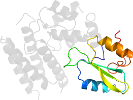
Domain d3vrqb2: 3vrq B:234-320 [250669]
Other proteins in same PDB: d3vrqa1, d3vrqa3, d3vrqb1, d3vrqb3
automated match to d1b47a3
complexed with ca; mutant
Details for d3vrqb2
PDB Entry: 3vrq (more details), 2.39 Å
PDB Description: Crystal structure of the tyrosine kinase binding domain of Cbl-c (PL mutant)
PDB Compounds: (B:) Signal transduction protein CBL-CSCOPe Domain Sequences for d3vrqb2:
Sequence; same for both SEQRES and ATOM records: (download)
>d3vrqb2 d.93.1.0 (B:234-320) automated matches {Human (Homo sapiens) [TaxId: 9606]}
nhpgymafltydevqerlqacrdkpgsyifrlsctrlgqwaigyvssdgsilqtipankp
lsqvllegqkdgfylypdgkthnpdlt
SCOPe Domain Coordinates for d3vrqb2:
Click to download the PDB-style file with coordinates for d3vrqb2.
(The format of our PDB-style files is described here.)
(The format of our PDB-style files is described here.)
Timeline for d3vrqb2:
- d3vrqb2 first appeared in SCOPe 2.04
- d3vrqb2 appears in SCOPe 2.06
- d3vrqb2 appears in the current release, SCOPe 2.08

 View in 3D