Lineage for d3nfua1 (3nfu A:1-132)
- Root: SCOPe 2.05

Class d: Alpha and beta proteins (a+b) [53931] (381 folds) 
Fold d.54: Enolase N-terminal domain-like [54825] (1 superfamily)
beta(3)-alpha(3); meander and up-and-down bundleSuperfamily d.54.1: Enolase N-terminal domain-like [54826] (2 families) 
Family d.54.1.0: automated matches [227195] (1 protein)
not a true familyProtein automated matches [226922] (83 species)
not a true protein
Species Chromohalobacter salexigens [TaxId:290398] [232962] (8 PDB entries) 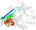
Domain d3nfua1: 3nfu A:1-132 [247901]
Other proteins in same PDB: d3nfua2, d3nfub2
automated match to d3mznb1
complexed with gol, mg, so4
Details for d3nfua1
PDB Entry: 3nfu (more details), 1.94 Å
PDB Description: crystal structure of probable glucarate dehydratase from chromohalobacter salexigens dsm 3043 complexed with magnesium
PDB Compounds: (A:) glucarate dehydrataseSCOPe Domain Sequences for d3nfua1:
Sequence; same for both SEQRES and ATOM records: (download)
>d3nfua1 d.54.1.0 (A:1-132) automated matches {Chromohalobacter salexigens [TaxId: 290398]}
lfpkitkmnvvpvagedgfllnlsgghepwfircvlvledesgnrgvgeipssegilngl
ekcrslvegarvnevkqvlsrargllaqggpeergrqtfdlrvavhvitaiesalfdlfg
qalgmpvadllg
SCOPe Domain Coordinates for d3nfua1:
Click to download the PDB-style file with coordinates for d3nfua1.
(The format of our PDB-style files is described here.)
(The format of our PDB-style files is described here.)
Timeline for d3nfua1:
- d3nfua1 first appeared in SCOPe 2.04
- d3nfua1 appears in SCOPe 2.06
- d3nfua1 appears in the current release, SCOPe 2.08

 View in 3D


