Lineage for d2vura1 (2vur A:41-178)
- Root: SCOPe 2.05

Class d: Alpha and beta proteins (a+b) [53931] (381 folds) 
Fold d.92: Zincin-like [55485] (2 superfamilies)
contains mixed beta sheet with connection over free side of the sheet
Superfamily d.92.2: beta-N-acetylhexosaminidase-like domain [55545] (4 families) 
contains similar fold but lacks its catalytic centre
Family d.92.2.3: Hyaluronidase N-terminal domain-like [143618] (2 proteins) 
Protein Hyaluronidase N-terminal domain [143619] (1 species) Species Clostridium perfringens [TaxId:1502] [143620] (7 PDB entries)
Uniprot Q8XL08 41-178
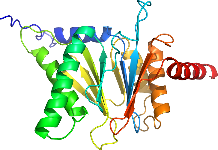
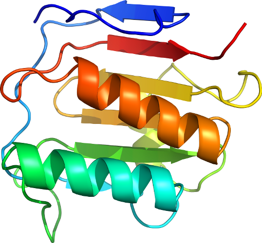
Domain d2vura1: 2vur A:41-178 [206488]
Other proteins in same PDB: d2vura2, d2vura3, d2vurb2, d2vurb3
automated match to d2cbia3
complexed with so4, yx1
Details for d2vura1
PDB Entry: 2vur (more details), 2.2 Å
PDB Description: chemical dissection of the link between streptozotocin, o-glcnac and pancreatic cell death
PDB Compounds: (A:) O-GlcNAcase nagJSCOPe Domain Sequences for d2vura1:
Sequence; same for both SEQRES and ATOM records: (download)
>d2vura1 d.92.2.3 (A:41-178) Hyaluronidase N-terminal domain {Clostridium perfringens [TaxId: 1502]}
vlvpnlnptpenlevvgdgfkitssinlvgeeeadenavnalrefltannieinsendpn
sttliigevdddipeldealngttaenlkeegyalvsndgkiaiegkdgdgtfygvqtfk
qlvkesnipevnitdypt
SCOPe Domain Coordinates for d2vura1:
Click to download the PDB-style file with coordinates for d2vura1.
(The format of our PDB-style files is described here.)
(The format of our PDB-style files is described here.)
Timeline for d2vura1:
- d2vura1 first appeared in SCOPe 2.03
- d2vura1 appears in SCOPe 2.04
- d2vura1 appears in SCOPe 2.06
- d2vura1 appears in the current release, SCOPe 2.08
 View in 3D