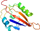
Lineage for d4hoib1 (4hoi B:28-137)
- Root: SCOPe 2.08

Class d: Alpha and beta proteins (a+b) [53931] (396 folds) 
Fold d.110: Profilin-like [55769] (10 superfamilies)
core: 2 alpha-helices and 5-stranded antiparallel sheet: order 21543; 3 layers: alpha/beta/alpha
Superfamily d.110.3: PYP-like sensor domain (PAS domain) [55785] (8 families) 
alpha-beta(2)-alpha(2)-beta(3)
Family d.110.3.0: automated matches [191387] (1 protein)
not a true family
Protein automated matches [190492] (24 species)
not a true protein
Species Mouse (Mus musculus) [TaxId:10090] [196720] (2 PDB entries) 
Domain d4hoib1: 4hoi B:28-137 [202454]
Other proteins in same PDB: d4hoia2, d4hoib2
automated match to d4hoia_
complexed with so4
Details for d4hoib1
PDB Entry: 4hoi (more details), 1.85 Å
PDB Description: Crystal structure of PAS domain from the mouse EAG1 potassium channel
PDB Compounds: (B:) Potassium voltage-gated channel subfamily H member 1SCOPe Domain Sequences for d4hoib1:
Sequence; same for both SEQRES and ATOM records: (download)
>d4hoib1 d.110.3.0 (B:28-137) automated matches {Mouse (Mus musculus) [TaxId: 10090]}
tnfvlgnaqivdwpivysndgfcklsgyhraevmqkssacsfmygeltdkdtvekvrqtf
enyemnsfeilmykknrtpvwffvkiapirneqdkvvlflctfsditafk
SCOPe Domain Coordinates for d4hoib1:
Click to download the PDB-style file with coordinates for d4hoib1.
(The format of our PDB-style files is described here.)
(The format of our PDB-style files is described here.)
Timeline for d4hoib1:
- d4hoib1 first appeared in SCOPe 2.03, called d4hoib_
- d4hoib1 appears in SCOPe 2.07
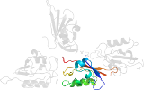
 View in 3D View in 3DDomains from other chains: (mouse over for more information) d4hoia1, d4hoia2, d4hoic_, d4hoid_ |


