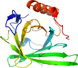
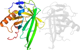
Lineage for d2acob1 (2aco B:11-175)
- Root: SCOPe 2.06

Class b: All beta proteins [48724] (177 folds) 
Fold b.60: Lipocalins [50813] (1 superfamily)
barrel, closed or opened; n=8, S=12; meander
Superfamily b.60.1: Lipocalins [50814] (10 families) 
bind hydrophobic ligands in their interior
Family b.60.1.1: Retinol binding protein-like [50815] (22 proteins)
barrel, closed; n=8, S=12, meander
Protein automated matches [190163] (13 species)
not a true protein
Species Escherichia coli [TaxId:562] [187394] (1 PDB entry) 
Domain d2acob1: 2aco B:11-175 [162757]
Other proteins in same PDB: d2acoa2, d2acob2
automated match to d1qwda_
complexed with vca
Details for d2acob1
PDB Entry: 2aco (more details), 1.8 Å
PDB Description: Xray structure of Blc dimer in complex with vaccenic acid
PDB Compounds: (B:) Outer membrane lipoprotein blcSCOPe Domain Sequences for d2acob1:
Sequence; same for both SEQRES and ATOM records: (download)
>d2acob1 b.60.1.1 (B:11-175) automated matches {Escherichia coli [TaxId: 562]}
lestslykkagstpprgvtvvnnfdakrylgtwyeiarfdhrferglekvtatyslrddg
glnvinkgynpdrgmwqqsegkayftgaptraalkvsffgpfyggynvialdreyrhalv
cgpdrdylwilsrtptisdevkqemlavatregfdvskfiwvqqp
SCOPe Domain Coordinates for d2acob1:
Click to download the PDB-style file with coordinates for d2acob1.
(The format of our PDB-style files is described here.)
(The format of our PDB-style files is described here.)
Timeline for d2acob1:
- d2acob1 first appeared in SCOPe 2.01, called d2acob_
- d2acob1 was called d2acob_ in SCOPe 2.05
- d2acob1 appears in SCOPe 2.07
- d2acob1 appears in the current release, SCOPe 2.08

 View in 3D


