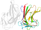
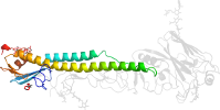
Lineage for d1vs8s1 (1vs8 S:1-110)
- Root: SCOPe 2.05

Class d: Alpha and beta proteins (a+b) [53931] (381 folds) 
Fold d.55: Ribosomal protein L22 [54842] (1 superfamily)
beta-alpha(3)-beta(2); 2 layers: alpha/beta; related to the enolase/MLE N-domain fold by a circular permutationSuperfamily d.55.1: Ribosomal protein L22 [54843] (1 family) 
some topological similarity to prokaryotic ribosomal protein L17Family d.55.1.1: Ribosomal protein L22 [54844] (1 protein) Protein Ribosomal protein L22 [54845] (5 species) 
Species Escherichia coli [TaxId:562] [160266] (29 PDB entries)
Uniprot P61175 1-110
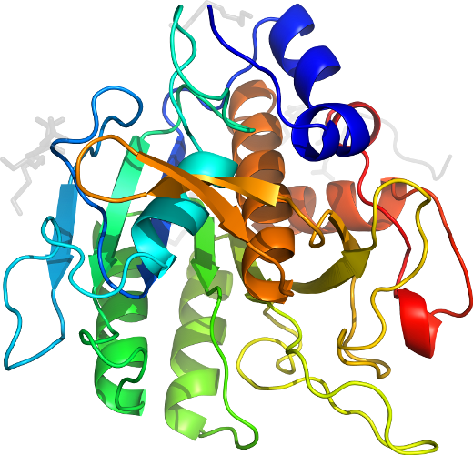
Domain d1vs8s1: 1vs8 S:1-110 [144513]
Other proteins in same PDB: d1vs801, d1vs811, d1vs821, d1vs831, d1vs841, d1vs8c1, d1vs8c2, d1vs8d1, d1vs8e1, d1vs8f1, d1vs8g1, d1vs8g2, d1vs8h1, d1vs8h2, d1vs8i1, d1vs8i2, d1vs8j1, d1vs8k1, d1vs8l1, d1vs8m1, d1vs8n1, d1vs8o1, d1vs8p1, d1vs8q1, d1vs8r1, d1vs8t1, d1vs8u1, d1vs8v1, d1vs8w1, d1vs8x1, d1vs8y1, d1vs8z1
complexed with mg
complexed with mg
Details for d1vs8s1
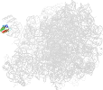
PDB Entry: 1vs8 (more details), 3.46 Å
PDB Description: Crystal structure of the bacterial ribosome from escherichia coli in complex with the antibiotic kasugamyin at 3.5a resolution. this file contains the 50s subunit of one 70s ribosome. the entire crystal structure contains two 70s ribosomes and is described in remark 400.
PDB Compounds: (S:) 50S ribosomal protein L22SCOPe Domain Sequences for d1vs8s1:
Sequence; same for both SEQRES and ATOM records: (download)
>d1vs8s1 d.55.1.1 (S:1-110) Ribosomal protein L22 {Escherichia coli [TaxId: 562]}
metiakhrharssaqkvrlvadlirgkkvsqaldiltytnkkaavlvkkvlesaianaeh
ndgadiddlkvtkifvdegpsmkrimprakgradrilkrtshitvvvsdr
SCOPe Domain Coordinates for d1vs8s1:
Click to download the PDB-style file with coordinates for d1vs8s1.
(The format of our PDB-style files is described here.)
(The format of our PDB-style files is described here.)
Timeline for d1vs8s1:
- d1vs8s1 first appeared in SCOP 1.75
- d1vs8s1 appears in SCOPe 2.04
- d1vs8s1 appears in SCOPe 2.06
- d1vs8s1 appears in the current release, SCOPe 2.08
 View in 3D View in 3DDomains from other chains: (mouse over for more information) d1vs801, d1vs811, d1vs821, d1vs831, d1vs841, d1vs8c1, d1vs8c2, d1vs8d1, d1vs8e1, d1vs8f1, d1vs8g1, d1vs8g2, d1vs8h1, d1vs8h2, d1vs8i1, d1vs8i2, d1vs8j1, d1vs8k1, d1vs8l1, d1vs8m1, d1vs8n1, d1vs8o1, d1vs8p1, d1vs8q1, d1vs8r1, d1vs8t1, d1vs8u1, d1vs8v1, d1vs8w1, d1vs8x1, d1vs8y1, d1vs8z1 |