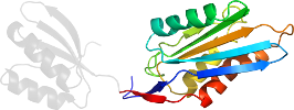
Lineage for d1vi7a1 (1vi7 A:3-137)
- Root: SCOP 1.75

Class d: Alpha and beta proteins (a+b) [53931] (376 folds) 
Fold d.14: Ribosomal protein S5 domain 2-like [54210] (1 superfamily)
core: beta(3)-alpha-beta-alpha; 2 layers: alpha/beta; left-handed crossover
Superfamily d.14.1: Ribosomal protein S5 domain 2-like [54211] (12 families) 

Family d.14.1.11: YigZ N-terminal domain-like [102772] (2 proteins)
modification of the common fold; contains extra alpha-beta unit after strand 2, the extra strand is inserted between strands 3 and 4
Protein Hypothetical protein YigZ, N-terminal domain [102773] (1 species) 
Species Escherichia coli [TaxId:562] [102774] (1 PDB entry)
two-domain structure is similar to the C-terminal region of EF-G (domains IV and V)
Domain d1vi7a1: 1vi7 A:3-137 [100740]
Other proteins in same PDB: d1vi7a2
structural genomics
Details for d1vi7a1
PDB Entry: 1vi7 (more details), 2.8 Å
PDB Description: crystal structure of an hypothetical protein
PDB Compounds: (A:) Hypothetical protein yigZSCOP Domain Sequences for d1vi7a1:
Sequence; same for both SEQRES and ATOM records: (download)
>d1vi7a1 d.14.1.11 (A:3-137) Hypothetical protein YigZ, N-terminal domain {Escherichia coli [TaxId: 562]}
lmeswlipaapvtvveeikksrfitmlahtdgveaakafvesvraehpdarhhcvawvag
apddsqqlgfsddgepagtagkpmlaqlmgsgvgeitavvvryyggillgtgglvkaygg
gvnqalrqlttqrkt
SCOP Domain Coordinates for d1vi7a1:
Click to download the PDB-style file with coordinates for d1vi7a1.
(The format of our PDB-style files is described here.)
(The format of our PDB-style files is described here.)
Timeline for d1vi7a1:
- d1vi7a1 first appeared in SCOP 1.67
- d1vi7a1 appears in SCOP 1.73
- d1vi7a1 appears in SCOPe 2.01
- d1vi7a1 appears in the current release, SCOPe 2.08

 View in 3D